Microscope Dentistry in Tirupati
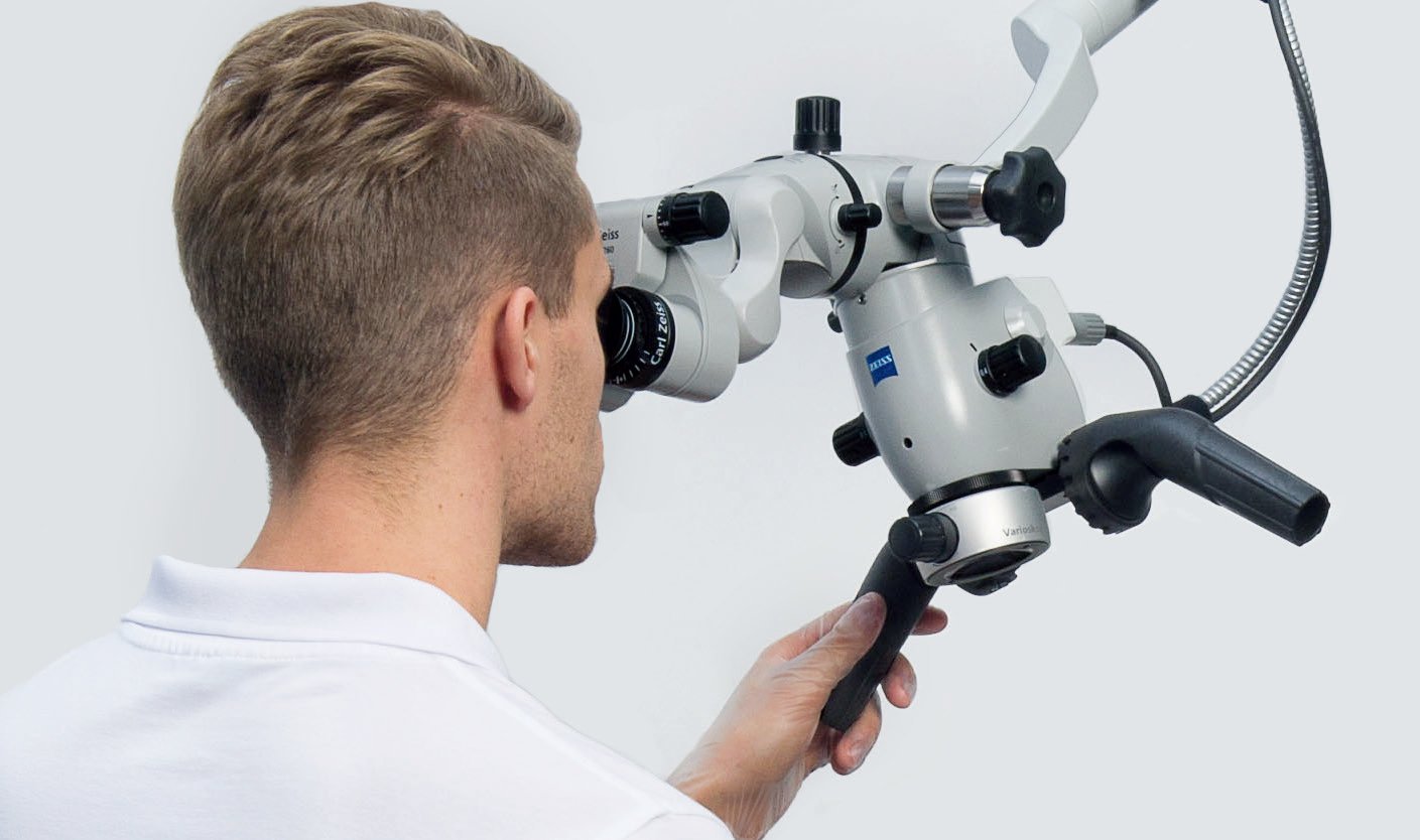
Tirupati's most precise dental care — powered by dental operating microscope with 25× magnification. See more, treat better, heal faster. Available only at Meghana Multi Speciality Dental Hospital.
Tirupati's most precise dental care — powered by dental operating microscope with 25× magnification. See more, treat better, heal faster. Available only at Meghana Multi Speciality Dental Hospital.
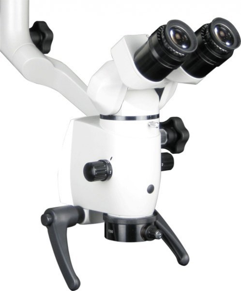
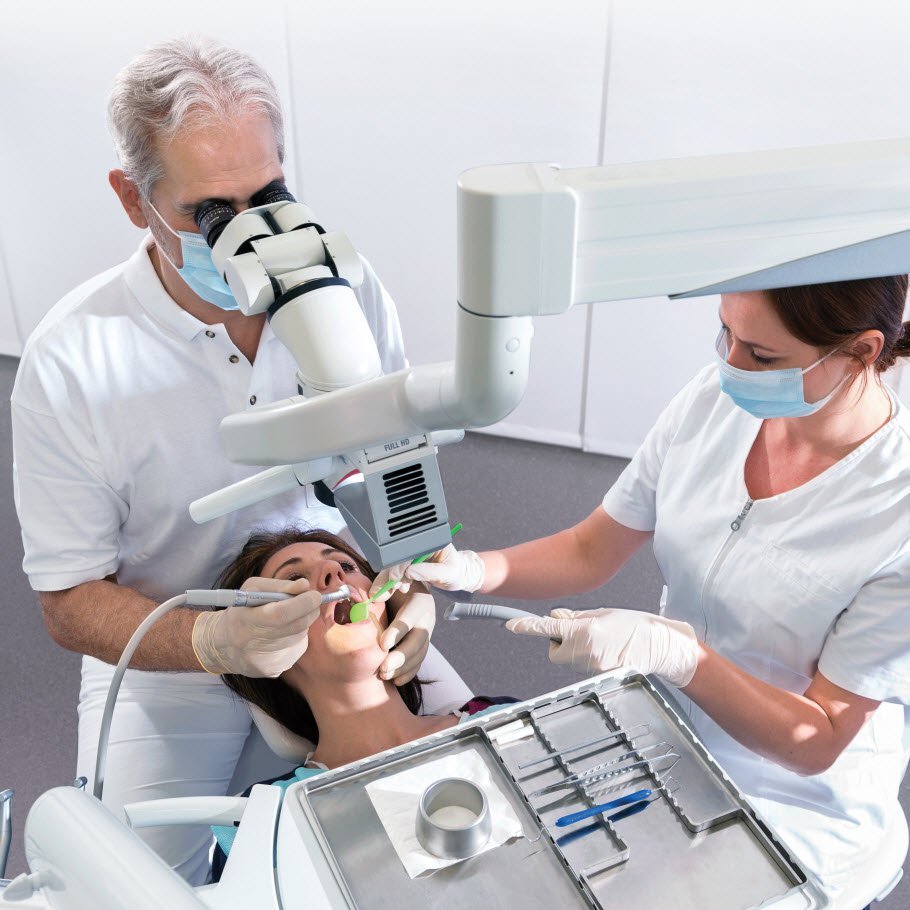
Meghana Multi Speciality Dental Hospital is the only dental clinic in Tirupati equipped with the dental operating microscope — the same gold-standard instrument used in leading dental universities worldwide. At up to 25× magnification, our specialist endodontist can visualise the entire root canal system, detect hairline fractures invisible to the naked eye, and perform microsurgical procedures with sub-millimetre precision. The result: treatments that are more thorough, more conservative, and significantly more successful than conventional approaches. If you have been told a tooth "cannot be saved" or a previous root canal has failed, microscope dentistry at Meghana Multi Speciality Dental Hospital offers a second chance backed by technology.

At 25× magnification every canal, crack, and anatomical detail is clearly visible — mistakes are not made in the dark.
Greater visibility means less healthy tooth structure is removed. We treat only what needs treating — nothing more.
Precise removal of infected tissue and clean canal preparation stimulates healthy tissue response and faster healing.
The microscope camera records the procedure. You can see exactly what the dentist sees — full transparency and accountability.
Peer-reviewed studies confirm microscope-guided endodontics achieves significantly higher success rates than conventional RCT.
When root canals are done right the first time under magnification, the need for costly and complex retreatments drops dramatically.
We begin with a detailed clinical examination and advanced 3D CBCT imaging where needed. This allows us to map the exact root anatomy, identify all canals, and plan the safest approach before touching the tooth.
Under the dental microscope at 8–25× magnification, the dentist creates a precisely sized access cavity. All canals — including calcified, curved, and accessory canals — are identified with certainty.
Rotary endodontic instruments guided by the microscope clean and shape each canal. Ultrasonic activation and antimicrobial irrigants flush out all infected tissue and bacteria from deep inside the root.
The prepared canals are filled with biocompatible gutta-percha under microscope confirmation. A perfect three-dimensional seal prevents reinfection and ensures long-term success.
After root canal treatment, the tooth is restored — typically with a zirconia crown — to protect it from fracture and return it to full biting function. The entire journey is documented.
Experience the most precise dental care in Tirupati. Whether it is your first root canal or a complex retreatment, our dental microscope with 25× magnification makes the difference.